By Meagan Goldman ’16
This article was inspired by Art of Science, an initiative to gather and exhibit scientific images from students, faculty, and staff. For more information, visit the Art of Science site.
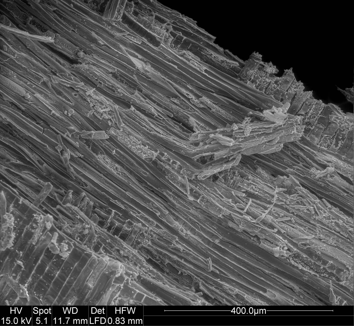
“It’s endlessly fun looking at these things,” says Nancy Piatczyc while enlarging a black and white image on her computer. As the image focuses, striations appear. Without context, it might be difficult to identify what it shows: a tiny fragment of wood magnified thousands of times by a scanning electron microscope (SEM). The wood is from a ship, likely British, that sank near the New York harbor around the time of the Revolutionary War. The SEM images will help biology professor Hank Art identify the wood from which the ship was built.
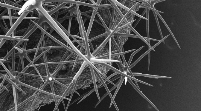
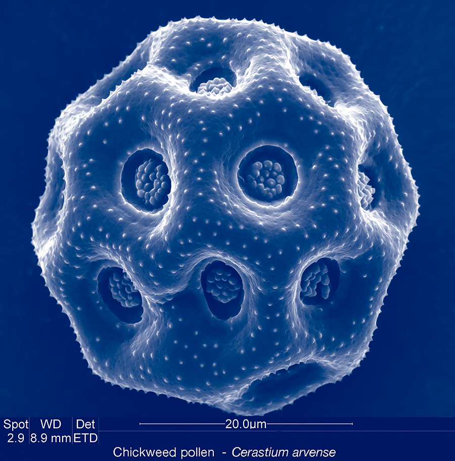
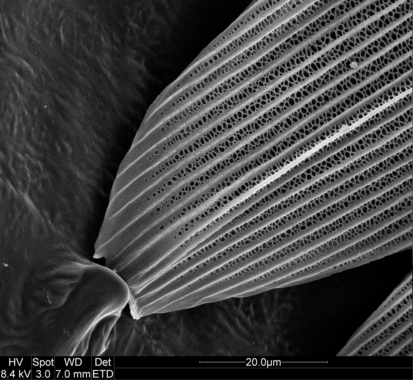
Examining wood from a sunken ship is one of the many varied projects of Nancy Piatczyc’s job. As the Williams electron microscopy technician, Piatczyc lives in a world that is normally invisible to the human eye. She works on projects for biology, chemistry, geosciences and physics, exploring the complex structures of living and non-living matter, from chickweed pollen (below) to cell nuclei and even individual atoms.
Born in Williamstown and raised in Stockbridge, Massachusetts, Piatczyc studied biology at Tufts University and electron microscopy at VA Medical Center in Albany, New York. She then worked as a technician at a dermatology lab in Ohio and a diagnostic tumor lab in Boston. When their daughter was one, she and her high school sweetheart husband decided to return to the Berkshires. Piatczyc frequently visited Williams to play with the electron microscope and eventually convinced the college to hire her as the technician. She has been here for twenty-nine years.
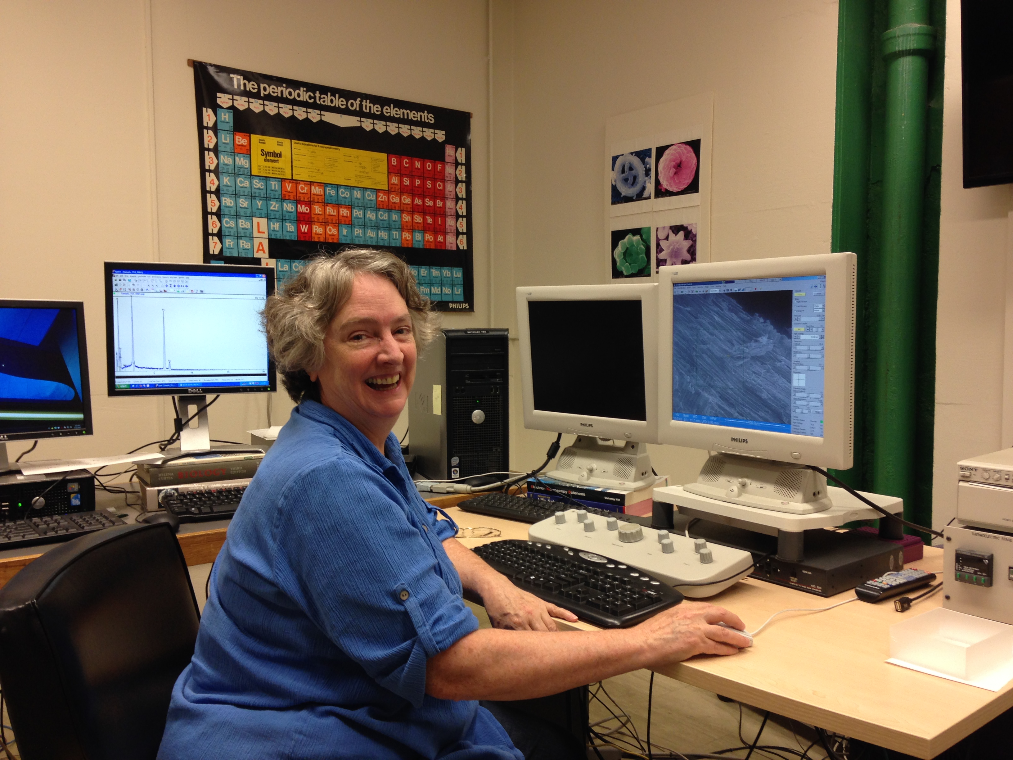
A patient and enthusiastic instructor, Piatczyc teaches microscopy to scientists of all ages. The facility is a valuable resource not only for Williams professors and students, but also for local middle school, high school, and community college students. They learn how to work three microscopes: the SEM, the transmission electron microscope (TEM), and the atomic force microscope (AFM). While the SEM creates images with depth and collects information about a sample’s composition, the TEM creates higher resolution images. The AFM generates topographic maps of samples, down to the atomic level.
In her free time, Piatzyc takes on miscellaneous projects like the sunken ship or works on projects of her own. She enjoys examining pollen and is collaborating with biology professor Joan Edwards to create a database of local plants. Her stunning images reveal the endless curiosity that drives her work.
“It’s rather amazing that things have such a beautiful structure at such a tiny scale.” she says. “I still find that amazing.”